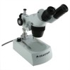
Celestron Advanced Stereo Microscope Microscope Basics - Page 14
Tungsten, Halogen, Fluorescent, Bright Field, Diffuse, Phase Contrast, Dark Field, Koehler
 |
View all Celestron Advanced Stereo Microscope manuals
Add to My Manuals
Save this manual to your list of manuals |
Page 14 highlights
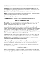
is needed then use the concave side. These methods are the least expensive illumination methods but it can be difficult to direct the light source for proper illumination. The more expensive and common illumination is by using built-in or attached light sources using bulbs or lamps that provide direct and intense illumination. These light sources can be from above the specimen or object which is used mainly with low power stereo microscopes and is called incident (reflected) light or from below a specimen (typically a slide specimen) which is light passing up through the specimen from inside the base and called transillumination (transmitted light). Lighting from both top and bottom at the same time can provide enough light for the most thick and irregular specimens. These illuminators may be of a fixed intensity, or of a variable intensity, which use a control knob (rheostat) to control the intensity of the light produced. Illumination lamps or bulbs come in various types: Tungsten - is an incandescent bulb filament which is the most common and least expensive. They give off a yellowish hue and give off moderate heat. They are typically 15-watt or 20-watt. Halogen - is a lamp which generally is the hottest light source for a microscope. The light is very bright, very white, and concentrated. The halogen type is more expensive than the tungsten. They are typically 15-watt or 20-watt. Fluorescent - is a lamp that is cool in temperature. The light is bright and white and very sharp while being comfortable to the eye. The fluorescent is great for observing live specimens. They are typically 5-watt to 10-watt and generate the same brightness as the tungsten or halogens do. They can be built in the base of a microscope or they can be attached (called a ring light) to observe from above. LED - these are light emitting diodes which provide a bright light source with virtually no heat. The white beam is brighter and cooler than the other illumination systems. They are typically battery operated and thus are cordless and great for outdoor use also. There are various forms of illumination produced by varying the amount of light or the quality of the light allowed to impinge on the specimen: Bright Field - this is the most fundamental and common form of lighting for microscopes. It is a highly directional and intense light source. Light aimed from beneath the stage through a condenser lens, through the specimen, through an objective lens, and through the eyepiece to the eye. Diffuse - this is where you place a ground glass, some translucent plastic, some opalescent material, or other similar material in front of the condenser (between the illuminator source and the condenser lens) and will cause the light of a bright field source to be scattered. Often this broadens the field illumination and brings subtle changes in the image. Phase Contrast - this form is used mainly because a large spectrum of living biological specimens (blood, tissue, and cultured cells), are virtually transparent or have poor contrast when observed with a bright field microscope. By utilizing a phase annulous (ring) mounted in the condensers front focal plane partially modulates the light ray bundles that pass through and around the specimen, where they are slowed ¼ wave, then are retarded another ¼ wave when they pass through the phase plate in the rear focal plane of the objective. This system also diminishes background light about 85% providing a darkened background to contrast with the illuminated structure of the phase object. While the affect diminishes the resolution of the image, it makes detail visible that one could not see without it. Dark Field - this form is a method to examine transparent or semi-transparent specimens which cannot be distinguished from the background. It shuts out background light and allows only scattered light to reach the specimen in order to heighten textural detail. Koehler - this form is a technique to optimize light quality and sharpness by aligning and adjusting each component of the optical system starting with a focusing illuminator. The light quality will be even and bright. The Koehler is the best form of illumination possible with a microscope and is offered on only the most expensive microscopes. Koehler Illumination 14
-
 1
1 -
2
-
3
-
4
-
5
-
6
-
7
-
8
-
 9
9 -
 10
10 -
 11
11 -
 12
12 -
 13
13 -
 14
14 -
 15
15 -
 16
16 -
 17
17 -
 18
18 -
 19
19
 |
 |
